.png)
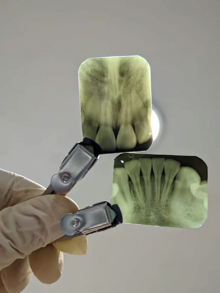
Every exam at Tanglewood starts with photos and x-rays reviewed before the doctor walks in. You see what we see. The conversation starts from a shared picture, not a verbal summary.

Early detection changes the treatment. A small cavity needs a filling. A cavity found six months later might need a crown.
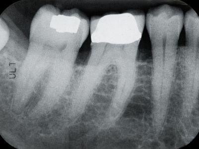
Cavities in their earliest stage, before they reach the inner tooth. Pearl flags them at a size most eyes miss.
Early bone loss around teeth or implants shows on x-rays long before it causes symptoms. We measure and track it.
Hairline cracks are invisible to the naked eye in a bright exam light. Intraoral photos under the right angle make them visible.
Thinning enamel from acid exposure or grinding. Visible in photos, trackable over time. Caught early it's manageable. Caught late it isn't.
Old fillings, crowns, and bonding that are beginning to break down before they actually fail. Replacing them on our schedule is far simpler than on an emergency.
Screened at every visit. Tissue changes that warrant attention are found when they're small. Most patients have no idea this is being done.
We let our patients do the talking.


Most offices rely on x-rays and the naked eye. We add two layers that find what those miss.
Pearl analyzes your x-rays before the doctor reviews them. It flags incipient decay, bone levels, and existing restorations at a detail level the naked eye misses. The doctor makes every clinical call. Pearl makes sure nothing gets overlooked.
We photograph the inside of your mouth at every exam. Fractures, enamel wear, tissue changes, failing restorations. You see what we're looking at, and we compare against your last visit so slow changes don't go unnoticed.
In order. No surprises.
Before the doctor comes in. Pearl flags areas of concern, highlights existing work, and notes anything worth a closer look.
Full set of photos of your teeth and tissue. Reviewed with you before the clinical exam begins. You see what we're looking at from the start.
The doctor examines each tooth, checks your bite, assesses your tissue, and reviews the Pearl findings and photos together. Everything is cross-referenced, not looked at in isolation.
We show you what we found, explain what it means, and tell you what we're monitoring versus what needs attention. You leave knowing where you stand, not waiting for a call.
Photos, x-rays, Pearl analysis, and clinical notes all filed. At your next exam, we compare against this visit. That's the unfair advantage of staying with the same practice over time.
For most patients, bitewing x-rays once a year and a full set every three to five years. We base the interval on your cavity history, bone health, and how much has changed since your last set.
It catches early-stage decay at a size and location that's easy for the human eye to overlook, particularly between teeth and near the gum line. The doctor still makes every clinical decision. Pearl flags what to look at twice.
Digital x-rays use a fraction of the radiation of traditional film. The dose is lower than what you receive on a short flight. We use lead aprons and take only what's clinically necessary.
Electric brushes perform better in clinical terms, but the best toothbrush is the one you actually use. If you do better with manual, use manual. We'd rather you brush consistently than perfectly.
A full set of x-rays, a complete series of intraoral photos, and a comprehensive exam. We'll build your baseline from scratch and tell you exactly what we find. No judgment on the gap.
We're fee-for-service, out-of-network with all major PPOs. We file your claim and reimbursement comes directly to you. Our fee schedule is on the website.
Whether you're looking to enhance your smile or simply maintain lifelong oral health, we’re here to guide you with expert care and honest conversations.
